
AI powered chemotherapy Monitoring
The OneTouch breast imaging system is an automated platform that combines photoacoustic imaging and ultrasound to capture detailed breast images while the patient is standing. This approach is designed to improve patient comfort, reduce operator variability, and provide fast, co-registered images without ionizing radiation or contrast agents.
By using ultrasound to show breast structure and photoacoustic imaging to highlight tumor-associated blood vessel patterns, OneTouch-PAT may help improve breast cancer diagnosis, tumor classification, and early prediction of treatment response.
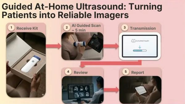
Guided at Home AI-Ultrasound
Breast ultrasound is an important imaging tool, especially for patients with dense breast tissue or suspicious breast findings. However, traditional ultrasound systems are often expensive, clinic-based, and dependent on trained operators, which limits their use for frequent or more accessible imaging. This project explores a low-cost, wireless handheld ultrasound approach supported by EchoWell Health-assisted image analysis to make breast imaging more scalable and patient-accessible. By testing deep learning models on both clinical and patient-acquired wireless ultrasound scans, this work evaluates whether suspicious breast regions can be meaningfully identified despite challenges such as lower image quality, scan variability, and differences from conventional ultrasound systems. Early results suggest that this framework may support future development of affordable, self-directed breast imaging and volumetric reconstruction.