Our Publications
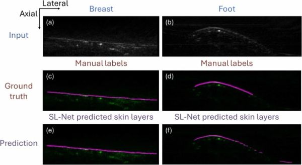
Enhanced clinical photoacoustic vascular imaging through a skin localization network and adaptive weighting
Photoacoustic tomography (PAT) is an emerging imaging modality with widespread applications in both preclinical and clinical studies. Despite its promising capabilities to provide high-resolution images, the visualization of vessels might be hampered by skin signals and attenuation in tissues. In this study, we have introduced a framework to retrieve deep vessels. It combines a deep learning network to segment skin layers and an adaptive weighting algorithm to compensate for attenuation. Evaluation of enhancement using vessel occupancy metrics and signal-to-noise ratio (SNR) demonstrates that the proposed method significantly recovers deep vessels across various body positions and skin tones. These findings indicate the method’s potential to enhance quantitative analysis in preclinical and clinical photoacoustic research.
Towards low-cost wireless AI-assisted breast tumor screening and volumetric freehand reconstruction
Breast ultrasound is valuable for patients with dense tissue and suspicious findings, but conventional systems remain limited by operator dependence, clinic-based infrastructure, and cost. These constraints restrict scalability for frequent imaging and deployment outside clinical settings. Advances in wireless handheld technology create opportunities for patient-performed imaging, but automated interpretation under constrained acquisition conditions remains unexplored. We present a low-cost, wireless, AI-assisted framework for self-directed breast ultrasound using a consumer-grade handheld probe with a mobile tablet interface. Rather than clinical screening performance, this work evaluates whether deep learning models trained on clinical data can generate meaningful predictions on patient-acquired wireless scans. Wireless imaging introduces technical challenges, including reduced channel count, simplified beamforming, lower signal-to-noise ratio, and acquisition variability, resulting in pronounced domain shift from conventional clinical data. We evaluated ResNet50, ResNeXt, and VGG16 for frame-level classification using over 5,000 frames from public datasets, institutional imaging, and wireless recordings, labeled as suspicious or non-suspicious. ResNeXt achieved the most stable performance (88.3% accuracy, 0.9692 AUC-PR). Applied to patient-acquired wireless cine loops from predefined freehand trajectories, model outputs produced localized suspicious prediction clusters showing qualitative spatial agreement with documented lesion locations. We also present preliminary IMU-based pose estimation toward volumetric reconstruction. This work demonstrates that AI-assisted interpretation can enable scalable, low-cost ultrasound workflows and informs engineering development of patient-accessible imaging systems.
Breast cancer imaging and neoadjuvant chemotherapy response prediction using the OneTouch photoacoustic imaging system
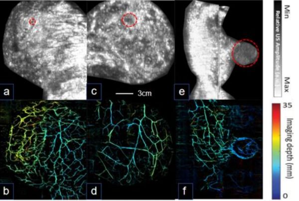
We present the OneTouch breast imaging system, an automated platform that simultaneously acquires co-registered photoacoustic (PA) and ultrasound (US) images of the breast in a standing position. This upright scanning approach aligns with conventional mammography workflows and improves patient comfort while reducing operator variability and exam setup time. The system provides three-dimensional US images of breast morphology and spatially matched PA images highlighting tumor-associated vascular features, without the use of contrast agents or ionizing radiation. In this study, we demonstrate the system’s clinical potential through representative imaging cases from patients with various breast cancer molecular subtypes, including luminal A, luminal B, and triple-negative tumors. These cases show subtype-associated variations in vascular architecture, which may enhance tumor classification beyond conventional imaging. In addition to diagnostic applications, we investigated whether baseline PA imaging could help predict response to neoadjuvant chemotherapy. Among four patients who completed treatment, we found that pre-treatment PA-derived vascular features—specifically endpoint ratio, and maximum branch length—differed between patients who achieved pathological complete response (pCR) and those who did not. These preliminary findings highlight the potential of OneTouch-PAT as a noninvasive tool for breast cancer diagnosis and early response prediction.
OneTouch Automated Photoacoustic and Ultrasound Imaging of Breast in Standing Pose
We developed an automated photoacoustic and ultrasound breast tomography system that images the patient in the standing pose. The system, named OneTouch-PAT, utilized linear transducer arrays with optical-acoustic combiners for effective dual-modal imaging. During scanning, subjects only need to gently attach their breasts to the imaging window, and co-registered three-dimensional ultrasonic and photoacoustic images of the breast can be obtained within one minute. Our system has a large field of view of 17 cm by 15 cm and achieves an imaging depth of 3 cm with sub-millimeter resolution. A three-dimensional deep-learning network was also developed to further improve the image quality by improving the 3D resolution, enhancing vasculature, eliminating skin signals, and reducing noise. The performance of the system was tested on four healthy subjects and 61 patients with breast cancer. Our results indicate that the ultrasound structural information can be combined with the photoacoustic vascular information for better tissue characterization. Representative cases from different molecular subtypes have indicated different photoacoustic and ultrasound features that could potentially be used for imaging-based cancer classification. Statistical analysis among all patients indicates that the regional photoacoustic intensity and vessel branching points are indicators of breast malignancy. These promising results suggest that our system could significantly enhance breast cancer diagnosis and classification.

Dual-Modal Photoacoustic and Ultrasound Imaging: From Preclinical to Clinical Applications
Photoacoustic imaging is a novel biomedical imaging modality that has emerged over the recent decades. Due to the conversion of optical energy into the acoustic wave, photoacoustic imaging offers high-resolution imaging in depth beyond the optical diffusion limit. Photoacoustic imaging is frequently used in conjunction with ultrasound as a hybrid modality. The combination enables the acquisition of both optical and acoustic contrasts of tissue, providing functional, structural, molecular, and vascular information within the same field of view. In this review, we first described the principles of various photoacoustic and ultrasound imaging techniques and then classified the dual-modal imaging systems based on their preclinical and clinical imaging applications. The advantages of dual-modal imaging were thoroughly analyzed. Finally, the review ends with a critical discussion of existing developments and a look toward the future.
Photoacoustic Dual-Scan Mammoscope: Results From 38 Patients
We have developed a photoacoustic-based imaging system, the dual-scan mammoscope (DSM), that combines optical contrasts with acoustic detection, to obtain the angiographic features in human breast. In this study, we investigated whether the system can differentiate malignant tumor and healthy breast. We have imaged 38 patients with various tumor types and compared results of tumor-bearing breast with healthy breast for each patient. We also compared the photoacoustic and ultrasound imaging results with clinical US. Vascular features in and around the tumor mass were visualized. We found that tumor-bearing breast contained vessels of larger caliber and exhibited stronger variations in the background signals than those in the contralateral healthy breasts. Preliminary data on photoacoustic and ultrasound images also indicate that the technique has potential in differentiating different tumor types. Overall, our results indicate that combining photoacoustic and ultrasound images can improve breast cancer screening. .

Volumetric Tri-Modal Imaging with Combined Photoacoustic, Ultrasound, and Shear Wave Elastography
Photoacoustic imaging is a hybrid imaging approach that combines the advantages of optical and ultrasonic imaging in one modality. However, for comprehensive tissue characterization, optical contrast alone is not always sufficient. In this study, we combined photoacoustic imaging with high-resolution ultrasound and shear wave elastography. The multi-modal system can calculate optical absorption, acoustic reflection, and stiffness volumetrically. We constructed a multi-modal phantom with contrast for each imaging modality to test the system’s performance. Experimental results indicate that the system successfully visualizes the embedded structures. We envision that the system will lead to more comprehensive tissue characterization for cancer screening and diagnosis.
Second-Generation Dual Scan Mammoscope With Photoacoustic, Ultrasound, and Elastographic Imaging Capabilities
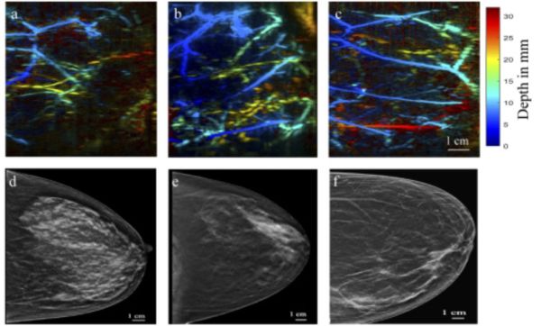
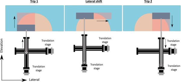
We recently developed the photoacoustic dual-scan mammoscope (DSM), a system that images the patient in standing pose analog to X-ray mammography. The system simultaneously acquires three-dimensional photoacoustic and ultrasound (US) images of the mildly compressed breast. Here, we describe a second-generation DSM (DSM-2) system that offers a larger field of view, better system stability, higher ultrasound imaging quality, and the ability to quantify tissue mechanical properties. In the new system, we doubled the field of view through laterally shifted round-trip scanning. This new design allows coverage of the entire breast tissue. We also adapted precisely machined holders for the transducer-fiber bundle sets. The new holder increased the mechanical stability and facilitated image registration from the top and bottom scanners. The quality of the US image is improved by increasing the firing voltage and the number of firing angles. Finally, we incorporated quasi-static ultrasound elastography to allow comprehensive characterization of breast tissue. The performance of the new system was demonstrated through in vivo human imaging experiments. The experimental results confirmed the capability of the DSM-2 system as a powerful tool for breast imaging.

Review of Methods to Improve the Performance of Linear Array-based Photoacoustic Tomography
As a hybrid imaging modality that combines optical excitation with acoustic detection, photoacoustic tomography (PAT) has become one of the fastest growing biomedical imaging modalities. Among various types of transducer arrays used in a PAT system configuration, the linear array is the most commonly utilized due to its convenience and low-cost. Although linear array-based PAT has been quickly developed within the recent decade, there are still two major challenges that impair the overall performance of the PAT imaging system. The first challenge is that the three-dimensional (3D) imaging capability of a linear array is limited due to its poor elevational resolution. The other challenge is that the geometrical shape of the linear array constrains light illumination. To date, substantial efforts have been made to address the aforementioned challenges. This review will present current technologies for improving the elevation resolution and light delivery of linear array-based PAT systems.

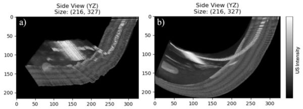
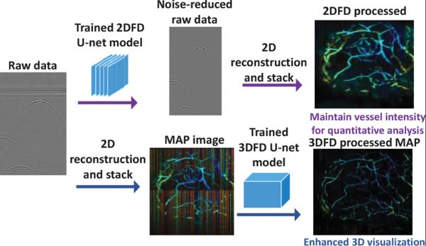
Deep Learning Enhanced Volumetric Photoacoustic Imaging of Vasculature in Human
The development of high-performance imaging processing algorithms is a core area of photoacoustic tomography. While various deep learning based image processing techniques have been developed in the area, their applications in 3D imaging are still limited due to challenges in computational cost and memory allocation. To address those limitations, this work implements a 3D fully-dense (3DFD) U-net to linear array based photoacoustic tomography and utilizes volumetric simulation and mixed precision training to increase efficiency and training size. Through numerical simulation, phantom imaging, and in vivo experiments, this work demonstrates that the trained network restores the true object size, reduces the noise level and artifacts, improves the contrast at deep regions, and reveals vessels subject to limited view distortion. With these enhancements, 3DFD U-net successfully produces clear 3D vascular images of the palm, arms, breasts, and feet of human subjects. These enhanced vascular images offer improved capabilities for biometric identification, foot ulcer evaluation, and breast cancer imaging. These results indicate that the new algorithm will have a significant impact on preclinical and clinical photoacoustic tomography.

Photoacoustic Imaging of Breast Cancer: A Mini Review of System Design and Image Features
Breast cancer is one of the leading causes for cancer related deaths in women, and early detection is extremely important to improve survival rates. Currently, x-ray mammogram is the only modality for mass screening of asymptomatic women. However, it has decreased sensitivity in radiographically dense breasts, which is also associated with a higher risk for breast cancer. Photoacoustic (PA) imaging is an emerging modality that enables deep tissue imaging of optical contrast at ultrasonically defined spatial resolution, which is much higher than that can be achieved in purely optical imaging modalities. Because of high optical absorption from hemoglobin molecules, PA imaging can map out hemo distribution and dynamics in breast tissue and identify malignant lesions based on tumor associated angiogenesis and hypoxia. We review various PA breast imaging systems proposed over the past few years and summarize the PA features of breast cancer identified in these systems.

Dual Scan Mammoscope (DSM)—A New Portable Photoacoustic Breast Imaging System With Scanning in Craniocaudal Plane
We present a new photoacoustic tomography system that provides visualization of angiographic features in a human breast with mammogram-like images. Methods: The system images a mildly compressed breast, from both top and bottom, using two 128-element, 2.25 MHz linear transducer arrays and line optical fiber bundles. The mild compression is achieved using plastic films, which is a more comfortable experience for the patient compared to rigid metal plates used in a traditional mammogram. Results: We could image a D cup-sized breast of 7 cm thickness within 1 minute and achieve a spatial resolution of around 1 mm in all directions. Conclusion: Our system possesses the benefits of portability, speedy scanning, and patient comfort. The craniocaudal-view images can be easily correlated with existing imaging modalities for data interpretation. Significance: Early cancer detection plays a critical role in overall cancer survival rate. Our system may address the limitations of mammogram and offer a radiation-free screening technique for patients with dense breasts.